
Where would you turn if your child
had a life-threatening brain tumor?

The Childhood Brain Tumor Foundation
presents
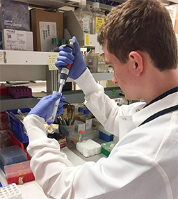
Expert VIDEO Updates on Childhood Brain Tumors

Patients and Families
CBTF works with patients and their families to help them find the information and resources needed to develop a strong understanding about childhood brain tumors.
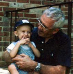
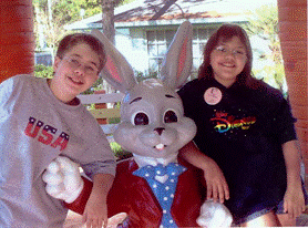
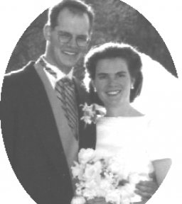
Personal Stories
CBTF families and patients share their personal journey and experiences.
INTERESTED IN SUPPORTING THE CHILDHOOD BRAIN TUMOR FOUNDATION?
Consider making a donation to support research for childhood brain tumors.